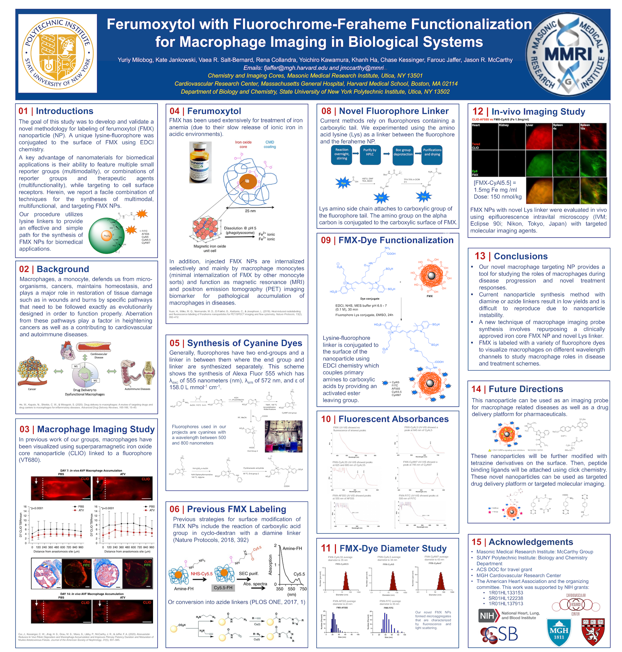
Ferumoxytol with Fluorochrome-Feraheme Functionalization for Macrophage Imaging in Biological Systems
Yuriy Milobog
Faculty: Lauren Endres
Macrophages are a type of immunine monocytes that serve roles such as protection against infections, restoration in wound healing, and homeostatic balance that occur in several intricately regulated pathways. When these pathways are disturbed, macrophages are proven to be pathogenic.
Macrophages have been connected to widespread disorders such vascular inflammation, autoimmunity, and a variety of malignancies. Identifying and distinguishing macrophages from other immune cells with macrophage-specific fluorophore-labeled contrast reagents are required to see and study these pathways.
Cyanine fluorophores with wavelengths of 600 to 700 nanometers are commonly employed in research; however, a current push for lower-wavelength fluorophores has led to the development of dyes with wavelengths around 500 nanometers, allowing for deeper tissue penetration.
Current chemical approaches for making macrophage-sensing nanoparticles are time-consuming, low-yielding, and frequently result in unstable nanoparticles that decompose. Using EDCI chemistry, our group developed a new approach that uses ferumoxytol nanoparticles (FMX) and a unique lysine-fluorophore linker attached to the nanoparticle surface.
Fluorescence and light scattering were used to characterize our new FMX nanoparticles, which generated microaggregates. Cyanine Cy-AL5, Cy-AM7, and a newly created Alexa Fluor 555 (AF555) fluorophore were produced and utilized in our work.
We used UV-VIS spectroscopy to validate FMX-fluorophore labeling, which was corroborated by the respectable absorbances of numerous fluorophores. We also measured the nanoparticle diameter to ensure that this novel nanoparticle is suitable for in-vivo macrophage imaging.
On in vivo near-infrared fluorescence (NIRF) imaging, FMX nanoparticles highlight macrophages and preferentially relocate to macrophage-rich tissues, according to preliminary findings.

